The cardio-respiratory model is based on the work of Dickinson, and comprises five functional units. These units are the:
1. Native Heart.
2. Native Lung.
3. Arterial Blood Pool.
4. Peripheral Tissue Pool.
5. Venous Blood Pool
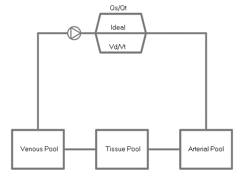
The model describes the passage of blood from the tissue pool through the venous pool and the native lung to the arterial pool - in an iterative fashion.
During a single iteration cycle, the blood is passed from one pool to the next in an amount which corresponds to the volume which has flowed during the iterative period. This incoming blood is then mixed perfectly with the blood in the existing pool and the new contents of oxygen, carbon dioxide and metabolic acids are calculated. When blood passes through the 'ideal compartment' of a lung pool, gas exchange occurs and oxygen and carbon dioxide diffuse across the alveolar:capillary membrane according to their pressure gradients. These lungs behave as 'Open Glottis' lungs which are exposed to atmospheric air (See: Hardman).
At the end of each iteration, oxygen is added to the tissue pool in an amount which corresponds to the amount which was acquired during transit through the lungs, and carbon dioxide is removed according to the amount which was expired in the ventilating gas.
Finally oxygen is removed from the tissue pool and carbon dioxide added to the pool according to the metabolic rate.
The native lung itself is modelled as as a classical 'Riley' three compartment structure.
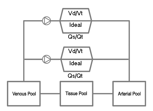
The model has been designed to allow for the inclusion of additional the lungs within the circuit. In particular, an artificial lung can be placed in parallel with the native lung (in order to simulate cardio-pulmonary bypass or veno-arterial ECMO), or in series with (before) the native lung (in order to simulate veno-venous ECMO). Alternatively, two native lungs (with variable parameters corresponding to the right and left lungs) can be placed in parallel with one another in order to simulate the physiology of differential lung ventilation (or one-lung anaesthesia).
All blood gas results are corrected to 37 C and reported at this temperature.
See:
Dickinson CJ. (1977). A Computer Model of Human Respiration/ MTP Press Limited. ISBN 0852001738
Hardman JG, Wills JS, Aitkenhead AR. Investigating hypoxemia during apnea: validation of a set of physiological models. Anesth Analg. 2000 Mar;90(3):614-8.
Riley RL and Cournand A. (1949) ‘Ideal’ Alveolar air and the analysis of ventilation-perfusion relationships in the lungs. J. Appl. Physiol., 1, 825-847